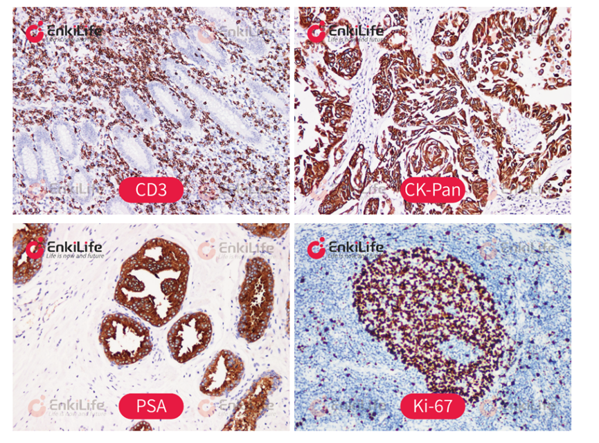
Immunohistochemistry (IHC) is a classical biological detection technology that integrates immunology, histochemistry, and microscopic imaging. Its core relies on the principle of antigen-antibody specific binding, combined with chemical colorimetric reactions, to achieve in situ qualitative, localization, and semi-quantitative analysis of target proteins within tissues and cells. It is one of the core technologies for life science research and clinical pathological diagnosis.
This technology primarily uses formalin-fixed paraffin-embedded (FFPE) tissue or frozen tissue sections as detection samples, with its core process including antigen-antibody reaction and signal amplification. First, the target antigen in the tissue section binds with a specific antibody (primary antibody); subsequently, a secondary antibody labeled with an enzyme or fluorophore binds to the primary antibody; finally, through chromogenic substrate or fluorescence excitation, the microscopic immune reaction is converted into a color or fluorescence signal visible under a microscope, thereby achieving visualization of the target molecule. For example, in the most widely used HRP-DAB system, HRP catalyzes DAB to form insoluble brown precipitate, allowing the location of the target antigen to be directly observed through a brightfield microscope. Depending on the connection method between the reporter enzyme and antibody, IHC can adopt detection modes such as direct method, indirect secondary antibody method, and polymer method. Among them, the polymer system achieves signal enhancement by enriching multiple reporter enzymes in the detection complex, providing a good balance between sensitivity, stability, and operational convenience, making it one of the common IHC detection methods currently.
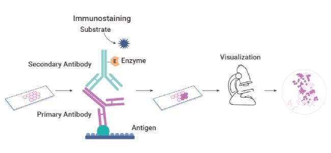
From the perspective of technology application, the value of IHC can be summarized as "specific recognition + in situ localization + visual detection". Antibodies provide specific recognition of target antigens, the detection system is responsible for converting this molecular recognition into observable signals, and tissue sections retain the spatial structure of cells and tissues. The combination of these three allows simultaneous observation of tissue morphology and target protein expression on the same section, thereby establishing the relationship between protein expression and cell types as well as tissue regions. For experiments that need to study protein localization, tissue heterogeneity, and disease-related tissue changes, IHC can provide spatial information that is difficult to obtain with simple protein quantification methods.
In the biological protein detection system, the core advantage of IHC lies in integrating specific recognition, in situ localization, and semi-quantitative analysis, achieving comprehensive detection from protein expression determination to tissue spatial information acquisition.
- Specific recognition: Accurately determine whether the target protein is expressed in the tissue to be tested, eliminate false positive interference from gene-level detection, and directly verify the true expression at the protein level.
- In situ localization: This is the core unique value of IHC. It can clarify the specific location of protein expression, distinguish nuclear, cytoplasmic, and membrane expression patterns, and simultaneously distinguish expression differences between tumor tissue, normal tissue, and stromal tissue, which is a core advantage that cannot be achieved by homogeneous sample detection technologies such as WB and qPCR.
- Semi-quantitative analysis: Relying on standardized pathological scoring systems, combining color intensity and positive cell proportion, graded quantification of protein expression levels can be used for inter-group difference comparison, drug efficacy evaluation, and disease grading analysis, fully meeting the data analysis requirements for scientific paper publication and pathological detection.
The core of immunohistochemistry is not merely "antibody binding to antigen"; more importantly, it is about how to convert this molecular recognition event occurring in the tissue microenvironment into stable, clear, and observable analyzable signals. A complete immunohistochemical detection system can typically be broken down into four key steps: antigen recognition, signal labeling, signal amplification, and signal presentation. Here, the primary antibody is responsible for recognizing the target antigen in the tissue, the detection system is responsible for converting the antigen-antibody binding into detectable signals, and different amplification systems determine whether the signal can be sufficiently enhanced. Finally, signals can be presented in brightfield form through chromogenic substrates such as DAB, or output in fluorescence form through fluorophores.
The direct detection method is a detection approach where enzyme molecules such as HRP, AP, or fluorophores are directly labeled on the primary antibody, without requiring secondary antibody participation. After the primary antibody specifically binds to the tissue target antigen, the colorimetric reaction can be directly initiated. This method has the fewest experimental steps and simple incubation layers, but this system has no signal amplification effect, overall detection sensitivity is relatively low, and the primary antibody activity after labeling is easily affected, with poor reagent versatility.
The indirect secondary antibody method is the basic indirect detection system of immunohistochemistry. It achieves specific binding through unlabeled primary antibody and tissue target antigen, then uses enzyme-labeled species-specific secondary antibody to bind to the primary antibody Fc segment, relying on the characteristic that a single primary antibody can bind multiple secondary antibodies to achieve signal amplification, and finally completes colorimetric imaging through enzymatic reaction. This system has simple overall operation process, strong reagent versatility, and stable experimental system, meeting the vast majority of detection needs, and is currently the most commonly used and most versatile basic detection system in immunohistochemistry experiments.
The avidin-biotin amplification system is a classical biotin-dependent detection system, mainly including two common methods, ABC and LSAB, achieving multi-level signal amplification through the ultra-high specific affinity between biotin and avidin. The ABC method is an early classical technique, where the experiment requires the use of biotinylated secondary antibody and avidin-biotin-reporter enzyme complex. Since avidin has tetravalent binding properties, it can form large molecular weight network complexes with extremely strong signal superposition effect and excellent detection sensitivity. The subsequently optimized LSAB method improved the core binding medium, relying on the one-to-one high-affinity specific binding between streptavidin and secondary antibody biotin to complete signal enrichment and amplification. This small complex has better tissue penetration ability, lower background interference, and higher detection sensitivity.
The polymer amplification system (EnVision system) is a non-biotin standardized detection system that avoids biotin interference, solving the high background problem of traditional biotin systems. Early polymer methods used dextran as the backbone, coupling multiple secondary antibodies and enzyme molecules to achieve signal amplification; new micro-polymer/dense polymer methods further optimized the structure, using smaller molecular weight detection complexes, effectively avoiding reagent aggregation, improving tissue penetration and staining uniformity, and further enhancing detection sensitivity. It is suitable for FFPE paraffin sections, frozen sections, and special tissues with biotin enrichment.
Traditional colorimetric IHC has advantages such as intuitive tissue structure, stable signal, and observability with brightfield microscopy, while DAB precipitation can better preserve the tissue localization information of target antigens, making it particularly suitable for tissue expression and localization analysis of single or few markers. However, as research questions extend from "whether a certain protein is expressed" to whether multiple proteins are co-expressed, which cell types different markers are localized in, and what spatial relationships exist between different cells, traditional monochromatic colorimetric IHC is gradually limited in multi-label detection and signal differentiation. In this context, fluorescence-based immunodetection technologies have further developed, providing more flexible technical means for multi-target co-detection, co-localization analysis, and tissue spatial information analysis.
Immunofluorescence converts antigen-antibody specific binding into detectable optical signals through fluorophores. Unlike colorimetric detection that relies on DAB to generate colored precipitates, immunofluorescence uses the excitation and emission characteristics of fluorescent molecules to achieve antigen localization and imaging. By selecting fluorophores with different emission wavelengths, multiple targets can be distinguished on the same tissue section, for example, using different colored fluorophores to label different proteins, combined with DAPI for nuclear staining, thereby achieving multi-marker detection and preliminary co-localization analysis. However, the signal intensity of conventional immunofluorescence mainly depends on the number of fluorophores carried by the detection system. For low-abundance antigens, the signal generated solely by fluorescently labeled secondary antibodies may be insufficient to form clear detection results. Therefore, to further improve the detectability of weak signal targets, fluorescence signal amplification technologies based on enzymatic catalysis have gradually been developed, among which TSA tyramide signal amplification is one of the representative technologies.
TSA (Tyramide Signal Amplification) is a highly sensitive signal amplification technology based on HRP enzymatic catalysis. Its core is not simply increasing the number of antibodies, but utilizing HRP to catalyze fluorophore-labeled tyramide in the presence of hydrogen peroxide to form reactive intermediates, which undergo in situ covalent deposition near the target antigen where HRP is located, thereby achieving high-density enrichment of local fluorescence signals. Compared with conventional immunofluorescence relying on fluorescently labeled antibodies carrying limited fluorescent molecules, TSA achieves signal accumulation through enzymatic catalysis, effectively improving the detection visibility of some low-abundance targets. Further combined with cyclic staining strategies, different targets can be sequentially detected according to the process of primary antibody recognition → HRP detection → TSA fluorescence deposition → antibody removal or inactivation → next target, retaining each target signal in its original spatial position, and ultimately achieving multi-marker detection, co-localization, and spatial relationship analysis on the same tissue section through multi-channel imaging.
| Catalog Number | Product Name |
|---|---|
| PA10014 | HRP Anti-Mouse IHC Detection System(DAB solution) |
| PA10015 | HRP Anti-Rabbit IHC Detection System(DAB solution) |
| RA10006 | HRP Anti-Mouse/Rabbit IHC Detection System(DAB solution) |
| PA10016 | Polymer-HRP Anti-Mouse IHC Detection System(DAB solution) |
| PA10017 | Polymer-HRP Anti-Rabbit IHC Detection System(DAB solution) |
| RA10007 | Polymer-HRP Anti-Mouse/Rabbit IHC Detection System(DAB solution) |
| PA10002 | IHC Double staining kit(Polymer-HRP)(DAB/AEC, For Mouse/Rabbit Primary Antibodies) |
| PA10020 | Rapid Ultra-Sensitive IHC Kit (pH 6.0) |
| PA10021 | Rapid Ultra-Sensitive IHC Kit (pH 9.0) |
| PA10001 | IHC Double staining kit(DAB/AEC, For Mouse/Rabbit Primary Antibodies) |
Experimental Results Display