
Antibody Labeling Kits: Unlocking the "Visualization" of Antibodies, a Research Essential
In life science research, clinical detection, and drug development, antibodies are indispensable core tools. They possess the extraordinary ability to precisely recognize and specifically bind antigens, serving as "precision probes" for researchers to track biomolecules, detect diseased cells, and screen targeted drugs. However, natural antibodies themselves have no luminescence, no color development, and no signal characteristics; their binding trajectories and results cannot be directly captured by the naked eye or conventional instruments. To make the antibody's working process "visible and quantifiable", antibody labeling must be performed to complete signal modification and functional enhancement of antibodies.
Core Principle: Equipping Antibodies with "Signal Tags"

Antibody labeling is essentially antibody conjugation technology, and antibody labeling kits are complete toolkits that standardize and simplify the complex conjugation chemistry workflow. Its core principle is very clear: the active labeling molecules in the kit can undergo mild covalent binding with specific amino acid residues on the antibody surface (lysine amino groups, cysteine sulfhydryl groups, sugar chain hydroxyl groups, etc.), stably connecting detectable signal tags to antibodies without destroying their antigen-binding activity, allowing the originally "invisible" antibodies to possess traceable and quantifiable detection signals.

Traditional manual labeling requires researchers to independently prepare reagents, control reaction conditions, and purify products. It not only requires a solid foundation in chemical experimentation, but also is highly prone to problems such as low labeling efficiency, antibody inactivation, and excessive product impurities. Commercial antibody labeling kits, however, have precisely proportioned and standardized packaged core components such as reaction buffers, activation reagents, labeling dyes, and termination solutions, optimizing reaction conditions and greatly lowering experimental barriers, allowing beginners to quickly complete high-quality labeling experiments.

Mainstream Types: Choose According to Research Scenarios

According to different labeling tags, mainstream antibody labeling kits on the market can be divided into four categories, covering the vast majority of basic research and clinical detection scenarios. Different types have different signal output methods and adapted experiments:

1. Fluorescent Labeling Kits
Fluorescent labeling kits are currently the most widely adopted and applicable labeling tools in life science imaging and cell analysis. Mainstream labeling substrates include classic fluorescent dyes such as FITC, Alexa Fluor series, Cy3, and Cy5. Their core principle is through specific covalent conjugation between dye active groups and antibody amino groups, allowing antibodies to carry specific fluorescent chromophores without interfering with antibody recognition sites. The biggest feature of these kits is substrate independence and direct imaging; after labeling, antibodies can stably emit specific fluorescent signals under excitation light of the corresponding wavelength. Among them, Alexa Fluor series dyes have strong photobleaching resistance and high fluorescence brightness, suitable for long-term microscopic observation; FITC is cost-effective and versatile, suitable for routine basic experiments; Cy series dyes have longer wavelengths and lower background interference. This type of kit is mainly adapted to experiments such as immunofluorescence staining (IF), flow cytometry (FCM), laser confocal scanning microscopy imaging, and tissue fluorescence localization, precisely achieving visualization and semi-quantitative analysis of target proteins in cells and membrane surface markers.
2. Enzyme Labeling Kits
Enzyme labeling kits are quantitative labeling tools using functional enzyme molecules as signal tags. The most commonly used labeling enzymes are horseradish peroxidase (HRP) and alkaline phosphatase (AP), which are also core supporting reagents for protein quantitative detection systems. Their labeling principle relies on mild cross-linking technology to stably conjugate enzyme proteins with antibodies, preserving both the antigen-specific binding ability of antibodies and the complete catalytic activity of enzymes. The two core enzymes have their own advantages: HRP has high sensitivity, fast reaction speed, and stable substrate chromogenic detection, adapting to most rapid quantitative experiments; AP has a milder catalytic reaction, wider signal linear range, and lower background color, suitable for high-precision, wide-range sample quantitative detection. Unlike the qualitative and semi-quantitative properties of fluorescent signals, enzyme-labeled antibodies can catalyze chromogenic substrates (TMB, pNPP, etc.) to undergo specific chromogenic reactions. The concentration of the colored product generated is highly linearly correlated with the target antigen content, and the absorbance value can be precisely read by a microplate reader to achieve precise quantification of samples. This type of labeling kit is mostly adapted to classical protein detection technologies such as ELISA, Western Blot protein blotting, and dot blot experiments. The products have extremely strong stability, small batch differences, and high experimental reproducibility. They are not only used for basic research protein expression quantification, but also core preparation tools for clinical in vitro diagnostic kit R&D and batch sample screening.
3. Biotin Labeling Kits
Biotin labeling kits belong to high-sensitivity auxiliary labeling tools, with small molecule biotin as the core label, relying on the natural super-specific binding system of "biotin-avidin" to achieve signal amplification. Biotin molecules are extremely small in volume; after labeling, they almost do not change the spatial conformation of antibodies, do not affect the binding efficiency of antibodies to antigens, and have the characteristics of controllable labeling density, extremely strong stability, and being less prone to inactivation. Different from directly labeled fluorescent and enzyme-labeled antibodies, biotin-labeled antibodies themselves have no dominant detection signal, but they can specifically bind to multivalent avidin/streptavidin. Through the cascade amplification effect of one avidin molecule binding multiple biotin-labeled antibodies and simultaneously carrying multiple signal groups, the detection signal intensity is multiplied, effectively solving the problems of weak signal, high background, and difficult detection of low-abundance target molecules. This type of kit is especially adapted to high-sensitivity demand scenarios such as trace sample detection, low-expression antigen localization, immunohistochemistry (IHC), in situ hybridization, and cell trace marker detection, widely applied in fields such as fine pathological detection and trace biomolecule mechanism research.
4. Specialized Functional Labeling Kits
Specialized functional labeling kits are precision labeling tools adapted to frontier scientific research and high-end R&D, mainly including subdivided types such as site-specific labeling, oligonucleotide labeling, and fluorescent quantum dot labeling, breaking through the technical shortcomings of traditional random labeling. Traditional kits mostly use random conjugation labeling, which may label antibody antigen-binding sites and slightly affect antibody activity; whereas site-specific labeling kits use patented targeted cross-linking technology to precisely target the antibody Fc constant region for site-specific labeling, completely avoiding the Fab antigen-binding region, fundamentally ensuring antibody activity and specificity, with labeling uniformity and experimental reproducibility far exceeding ordinary kits. Oligonucleotide labeling kits can conjugate specific nucleic acid sequence tags to antibodies, adapting to frontier technologies such as immune sequencing, single-cell multi-omics analysis, and high-throughput protein spectrum screening; quantum dot labeling kits possess both ultra-high fluorescence stability and multi-color imaging capabilities, suitable for multi-indicator simultaneous localization detection. These high-precision kits mainly serve frontier fields such as antibody-drug conjugate (ADC) R&D, single-cell sequencing, high-end proteomics research, and precision targeted drug screening, and are core supporting tools for high-end biomedicine R&D and top-tier mechanism research.
Core Advantages and Application Scenarios: Standardization Empowers Full-Field Scientific Research

Compared to the traditional method of manually preparing reagents and manual conjugation labeling, standardized antibody labeling kits have irreplaceable operational advantages, thoroughly solving the pain points of traditional labeling technology such as cumbersome operation, high barriers, low success rate, and large errors. They precisely proportion and standardize package all reagents including reaction buffer, activation reagent, labeling substrate, and preservation solution. The reaction system is mild and controllable, requiring experimenters to have no professional organic conjugation chemistry foundation; beginners can quickly complete labeling experiments through standardized workflows, greatly shortening the experiment exploration cycle and reducing trial-and-error costs. Meanwhile, the mild reaction system can maximally preserve the natural spatial structure and antigen-binding activity of antibodies. Labeled products have strong stability, can be stored long-term, and have extremely small batch-to-batch reagent differences, effectively avoiding manual operation errors.

1. Small Molecule Fluorescent Label Application Examples

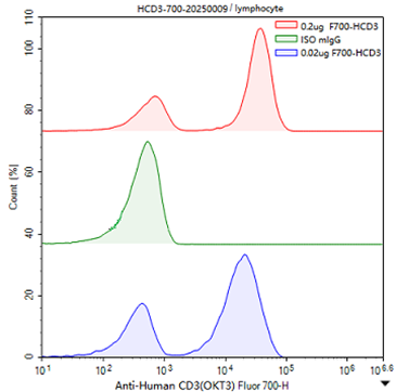
Small molecule fluorescent labels have rich categories and comprehensive spectral coverage, adapting to single-label, multi-label fluorescence imaging and flow detection, making them the mainstream labeling solution for basic immune experiments. Using Fluor700 small molecule fluorescent labeling kit (SKU: E80026) to label mouse anti-human CD3 (OKT3) antibody, applied to lymphocyte flow typing experiments. After loading, the negative background is clean, positive T cell populations are clearly separated, with no obvious diffuse signals, and typing results are precise and stable.
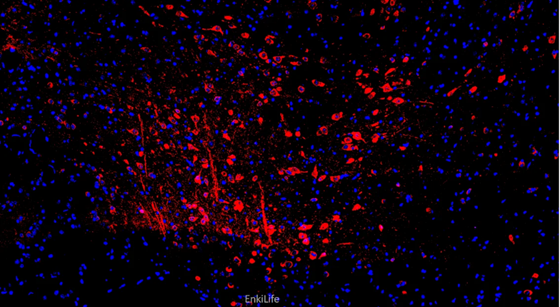
Using Fluor555 labeling kit (SKU: E80012p) to label UCH-L1/PGP9.5 rabbit polyclonal antibody, performing direct immunofluorescence staining on 4% paraformaldehyde-fixed mouse brain tissue sections at a 1:200 dilution ratio. Target protein localization is precise, fluorescent signals are uniform, and the distribution characteristics of target proteins in brain tissue can be clearly displayed.
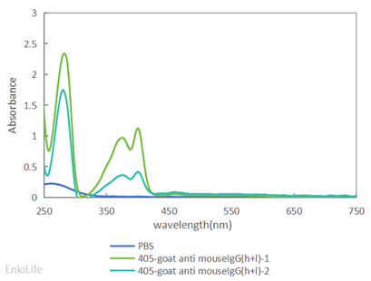
After Fluor405 labeling kit (SKU: E80010p) labels goat anti-mouse IgG antibody, the spectral absorption peak is regular and fluorescence output is stable, perfectly adapting to the blue channel compensation matching needs in multi-color experiments.
2. Tandem Protein Fluorescent Label Application Examples

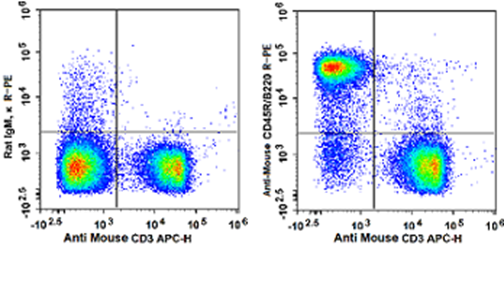
PE and APC series tandem fluorescent protein labels have the advantages of ultra-high fluorescence brightness and low cross-color, specifically adapted to multi-color flow cytometry detection and high-sensitivity cell typing experiments. Using R-PE high-brightness tandem dye (SKU: E80005p) to label rat anti-mouse CD45R antibody and APC fluorescent protein (SKU: E80006p) to label mouse CD3 antibody, conducting dual-indicator lymphocyte simultaneous detection. In the experiment, each fluorescence channel has high discrimination, no obvious spectral cross-talk, and positive cell populations are tightly clustered, efficiently achieving precise immune cell subtyping.
3. HRP Enzyme Label Application Examples

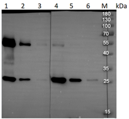
HRP enzyme labels have good chromogenic stability and excellent concentration linear response, making them core functional labels for protein blotting and quantitative immune detection. Using HRP labeling kit (SKU: E80004p) to respectively label goat anti-mouse IgG whole molecule antibody and F(ab)₂ fragment antibody, conducting WB verification experiments against mouse IgG samples at gradient concentrations of 500ng, 50ng, and 5ng. The results show that band gradients of samples at different concentrations are distinct, trace protein samples at ultra-high dilutions can still show clear chromogenic detection, with no miscellaneous bands, no high background color, and no false positive interference. This labeling system has extremely strong reproducibility, not only suitable for routine protein qualitative verification, but also meeting experimental needs for precise quantitative screening of large-batch ELISA samples.
Conclusion

Small antibody labeling kits, seemingly auxiliary tools in experiments, are actually key bridges connecting antibodies and detection signals. They popularize and standardize complex bioconjugation technologies, allowing researchers to focus on core topic research without getting tangled in tedious chemical experiment details. From basic protein detection to frontier antibody drug development, from laboratory research to clinical precision diagnosis, this "research essential" continues to escort technical breakthroughs and application implementation in the life sciences field.

EnkiLife covers fluorescent, enzyme, and biotin series labeling kits, adapting to research scenarios such as flow cytometry, IHC, basic immunology, and vaccine development. It also provides one-stop custom labeling outsourcing, with simple operation, high product activity, and stable batches, preferred by laboratory researchers.

   💬 WhatsApp